The NMR laboratory of the Structural Biology Platform is equipped with 3 NMR spectrometers:
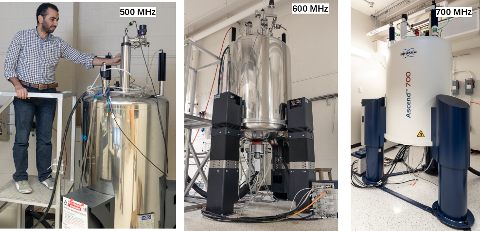

- A 500 MHz spectrometer, equipped with either a triple resonance (TXI) or quadrupole resonance (QXI) room temperature probe.
- A 600 MHz spectrometer, equipped with either cryogenic (TCI) or room temperature (TXI) triple resonance probe.
- A 700 MHz spectrometer, equipped with either a triple resonance (TXI) or double resonance broadboard (BBI) room temperature probe.
...
Each NMR spectrometer is composed of various parts that will be discussed below:
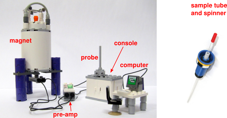

Source: LEGO ideas
...
Several software are in place in order to control the various pieces of the NMR spectrometer and analyze the data. The following programs are installed on the control computers:
| Name | Purpose | Command to launch |
|---|---|---|
| TopSpin | Main control and processing software | topspin |
| BSMS | Bruker Smart Magnet Control System | bsmsdisp (within TopSpin) |
| MICS | Magnet Information and Control System | mics (within TopSpin) |
| NMRPipe | Advanced processing scripts | nmrPipe |
NMR magnet
The magnet assembly (aka the can) is often the most impressive piece of equipment in a NMR laboratory due in part to the size of its cryostat. Depending on the field strength, they can easily reach two stories high (above 5 meters in height).
...
NMR magnets present numerous health hazards caused by the presence of strong magnetic fields. The strength of the magnetic field increases with the proximity to the core of the magnet. Each magnet in the NMR laboratory has its own particularities with respect to stray field. Do not cross the yellow plastic chain (located at the 5 Gauss (0.5 mT) line) before making sure you do not have anything metallic on you and in your pockets.
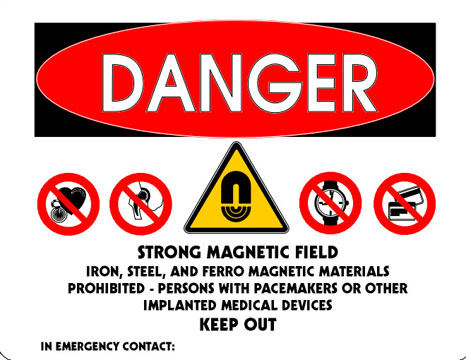

Rules to follow around the NMR magnets:
...
The NMR magnet is composed at is core of an active electromagnet created by winding several kilometres of superconducting wire kept below its critical temperature (resistance close to zero at near absolute zero) by immersion in liquid helium at 4.2 K. This is then kept cool using a jacket filled with liquid nitrogen at 77 K.


Source: Course Hero
...
Below is an example of the cross-section of a real magnet. It includes (2) the foil radiation isolation, (5) the liquid nitrogen cryostat, (10) the superconducting wire, (13) the room temperature bore tube and (20) the liquid helium cryostat. A list of all the different identified parts can be found here.
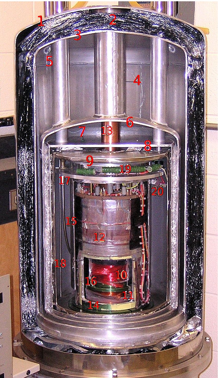

Source: H.A. Trujillo, Wilkes U.
...
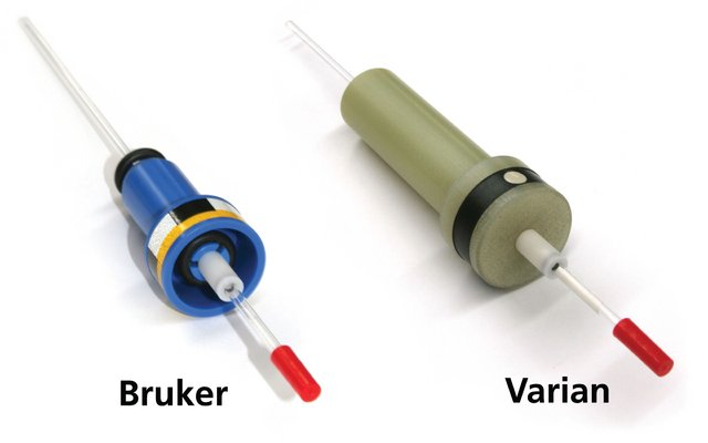
The sample has to be placed in a thin borosilicate glass wall tube of 5 mm in diameter rated at least to the proton frequency of the spectrometer used. Our probes can also accept Bruker-type Shigemi, 3 mm and shaped tubes. Then, the sample tube has to be inserted first into a spinner which is basically a sample holder. Make sure you use a Bruker spinner, not a Varian one. The difference is illustrated below.

POM (polyoxymethylene) blue plastic spinners are the standard spinners used for most bio-NMR experiments. However, if you need to run your experiment at temperatures between 0 and 5 °C, or above 80 °C, you will need to use a Kel-F (polychlorotrifluoroethylene) white spinner which is heavier and more resistant to high temperatures (do not exceed 150 °C!). Air flow from the BCU II will also need to be adjusted also.
It is also imperative that you use the sample depth gauge to ensure that your sample tube is set at the proper depth, as illustrated below. If you insert your tube too deep, it will hit the bottom of the probe insert and will likely break, resulting in a potential expensive cleaning and repair procedure. Alternatively, a sample that is not inserted enough will not be fully exposed to the coils of the probehead.

Source: Bruker - BSMS manual
...
Below is the general schematic of a NMR probe, including the position of the gradient coils, outside of the RF coils.
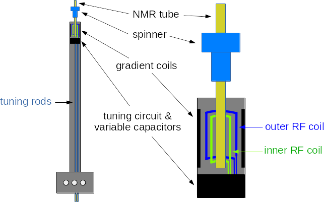

Source: UCSD Skaggs School of Pharmacy and Pharmaceutical Sciences NMR Facility
And a picture of the actual coils, if we were to remove the protective plastic:
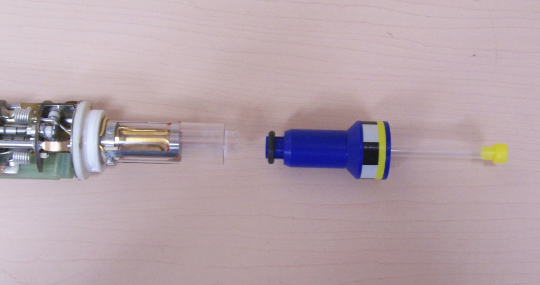

Source: H.A. Trujillo, Wilkes U.
...