Healthy cells look like this.
...
What is photo-toxicity?
Under appropriate temperature (37°C), humidity (hight) and atmosphere (5% CO2) it is possible to grow cells are growing in culturevitro.
Healthy cells
But looking at these Looking at cells under the microscope is not without riskimpact. This is similar to when you are exposing yourself to the sun light. If the sun is too strong and/or if you are exposed for a too long period you can have a bad sun burn...The The same apply applies to cells under a microscope. While transmitted light (bright-field, phase-contrast, DIC) does not affect cell biology, fluorescence excitation light can be deleter.
How does photo-toxicity look like?
Strong illumination light will eventually burn your cells. Adhering cells will retract, detach and eventually die. See the example below (Click on the image to see the movie). These are cells exposed to 1 second of 561nm 70mW of 550nm light every 5 minutes. The total movie is about 3h. The cells retract, detach and eventually die.
Click on the image to see the movie
Below This is another example of cells exposed to 1 second of 59mW of 405nm 395nm and 1s of 561nm 70mW of 550nm light every 5 minutes. The total duration is about 8h.
Click on the image to see the movie
How can I identify photo-toxicity has occured?
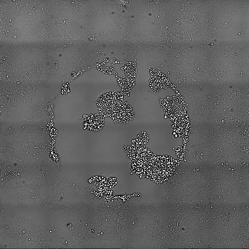
The best way to do this is to take a larger field of view image after your acquisition. Because photo-toxicity occurs only in the illuminated area, taking a picture of adjacent cells will provide with a very good (but not perfect) approach.
If you step back then you can see that the damaged are area is actually limited to the exposed area. Note: The recorded area is smaller than the exposed area.
Importantly this is not a perfect control. A more ideal control would be to have another dish with cells maintained in the same condition but without illumination. In fact in the image above we can't conclude whether or not the affected circular area has no impact on the surrounding cells. It is possible that cell death occurring under the illuminated field of view can produce and release molecules that will affect the surrounding area. Therefore the ideal control would be a totally separated dish.
What are the important factors when talking about photo-toxicity?
The amount of light, the wavelength, the surface illuminated, the duration of illumination, the repetition of the illumination.
...
The wavelength: The energy carried by laser light is dependant dependent of the wavelength. Shorter wavelength have more energy and are more deleterious
...
With a power meter you can also evaluate the amount of energy that your cells are confortable comfortable with.
To do this measure the power at the objective using your regular imaging settings.
...
Cells were imaged for 100ms with 561nm 70mW of 550nm light every 5 minutes. Cells are dividing faster than the effect of photo-toxicity that is occurring. Again here the best way to control is to take an overview image at the end of the acquisition and compare exposed cells to non-exposed cells.
Click on the image to see the movie


