Healthy cells look like this.
Under appropriate temperature (37°C), humidity (hight) and atmosphere (5% CO2) cells are growing in culture.
Looking at cells under the microscope is not without risk. This is similar to when you are exposing yourself to sun light. If the sun is too strong and/or you are exposed for too long you can have a bad sun burn...
The same apply to cells under a microscope.
These are cells exposed to 1 second of 561nm light every 5 minutes. The total movie is about 3h. The cells retract, detach and eventually die.
Click on the image to see the movie
This is another example of cells exposed to 1 second of 405nm and 1s of 561nm light every 5 minutes. The total duration is about 8h.
Click on the image to see the movie
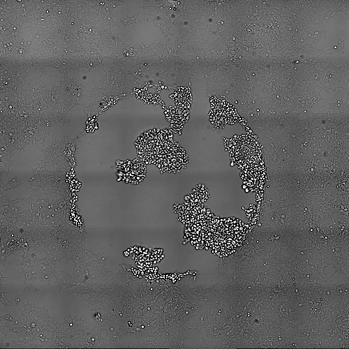
If you step back then you can see that the damaged are actually limited to the exposed area
What are the important factors when talking about photo-toxicity?
The amount of light, the wavelength, the surface illuminated, the duration of illumination, the repetition of the illumination.
Amount of light: If you shine a strong light vs a dim light
The wavelength: The energy carried by laser light is dependant of the wavelength. Shorter wavelength have more energy and are more deleterious
The area illuminated: Shining the same amount of light into a small area result in stronger damage
The duration of illumination: Shining light for 1 second is doing more damage than 10 ms
The repetition: Shining 10 ms 20 times per minute is doing more damage than 1 time per minute
Empirically you can determine if photo-toxicity occur by looking at your cells. IF your cells are not dividing, if they detach then you may have photo-toxicity.
It is always good to acquire a larger field of view of the recorded area to make sure no photo-toxicity has occurred.
With a power meter you can also evaluate the amount of energy that your cells are confortable with.
To do this measure the power at the objective using your regular imaging settings.
You will obtain a value in mW (which is Joule/second).
Then divide this value by the field of view of your objective. This will provide the irradiance expressed in mW/cm2.
Finding the irradiance that is stress free or your cells is key. To do this you can shine continuously a defined irradiance and observe your cells for several hours. If your cells do not display any sign of photo-toxicity you can increase the irradiance and continue until finding the maximum acceptable continuous irradiance.
This will provide a good idea of how strong are your cells.
Obviously there is some flexibility since you will likely not continuously expose your cells. So you may pass above the maximum acceptable continuous irradiance value, which will stress your cells but eventually they will recover. Only you can determine if this amount of stress is experimentally acceptable and not modify the biology of what you want to measure.
Here is another discrete example.
Cells were imaged for 100ms with 561nm light every 5 minutes. Cells are dividing faster than the effect of photo-toxicity that is occurring. Again here the best way to control is to take an overview image at the end of the acquisition and compare exposed cells to non-exposed cells.
Click on the image to see the movie


