To maximize the quality of the NMR data acquired with our spectrometers, it is key to correctly handle the sample throughout the process; from production to NMR data acquisition. The objective of this section is to propose general guidelines for sample preparation and initial data acquisition.
Quantity
Biological NMR is intrinsically poorly sensitive, and the intensity of the signal is proportional to the amount of material present in the observed volume. Thus, you should maximize the quantity of sample submitted for analysis, while minimizing the volume, in order to get the highest concentration as possible.
Volume and concentration
If you use a regular 5 mm NMR tube, a filling length of 40 mm is recommended, which corresponds to about 550 µl of sample in a 5 mm tube with walls 0.4 mm thick. If the volume is limited, 5 mm Shigemi (ie containing susceptibility-matched plugs), or 3 mm tubes (with slightly reduced sensitivity), can be used and will require less volume. Below is an illustration of the construction of a Shigemi tube:
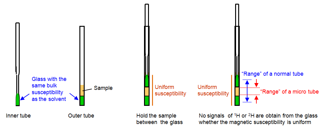
Source: JEOL
Summary of optimal volume and quatity for different NMR tubes:
| Type of tube | Volume, µl | Quantity, nmol | |
|---|---|---|---|
| at 100 µM | at 500 µM | ||
| 5 mm | 550 | 55 | 275 |
| 5 mm Shigemi | 350 | 40 | 200 |
| 3 mm | 160 | 20 | 100 |
Practically, on the instruments of the Structural Biology Platform, at least 100 µM of sample is sufficient for simple checks (1D and 2D correlation experiments), whereas concentrations beyond 0.5 mM should be attained in order to have good SNR on multi-dimensional experiments within acceptable experiment time. When the quantity and volume are limited, some options are available to obtain a good signal-to-noise ratio (SNR):
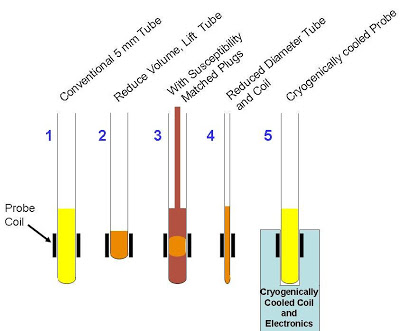
Source: UOttawa NMR facility blog
One thing to remember: we can increase the SNR by increasing the number of scans (ns), however the SNR increases as the square root of the number of scans recorded. So to get a similar SNR with a sample concentrated at 0.5 mM vs at 1.0 mM, 4 times more acquisition scans will be required (ie the experiment will take 4 times longer!).
SNR ∝ √(nb scans)
Stability
The sample submitted for NMR analysis has to be chemically stable over the course of the experiment and presence of contaminants (microbial, chemical, biochemical) could degrade it to a level beyond usability, rapidly, especially since most NMR recordings are done at, or near, room temperature. It is therefore imperative to evaluate the stability of the sample prior to the NMR experiments. This can easily be done by keeping an unlabeled sample at room temperature for 2 weeks and verifying its integrity before and after (SDS-PAGE, 1D NMR).
Additionally, the integrity of the sample should be assessed before and after long (more than 6 hours) experiments by running either a 1D proton spectrum, or, better for labeled samples, a 2D ^15^N-HSQC spectrum. If there is no difference in the spectra, no change in the chemical environment of the atoms has occurred, hence the sample remained identical (no degradation etc). Simply put:
identical spectra=identical sample
Sample buffer
Buffer pH
In terms of buffer, a slightly acidic environment (pH around 5.5 - 6.5) is typical for biological samples (proteins, DNA, RNA) in order to favor protonation of the exchangeable protons (amide (proteins) and imino (nucleic acids)). Above a pH of 7.5, it may be difficult to detect amide protons, more sample will be required, and can result in sample degradation.
Buffer ionic strength
Salt (typically in the form of NaCl or KCl) is generally used in sample buffers in order to prevent non specific ionic interactions and favor proper folding of the sample. For NMR analysis, ionic strength with a salt concentrations up to 500 mM is generally appropriate (below 150 mM being ideal). Higher salt concentrations complicate the tuning and matching of the probe and result in longer 90° pulse duration which may need power adjustments exceeding maximal permitted levels for a given probe. This is particularly true for the cryoprobe.
The case of the cryoprobe
When a cryogenic probe is used (as in the case of the TCI probe on our 600 MHz), salt concentrations below 100 mM are preferred in order to reduce signal degradation caused by the increased conductivity, which is a function of both the ion concentration and ion mobility .
This latter property may be modulated by using different compounds to prepare the sample buffer, and may even enable the use of high salt concentrations. Kelly et al describe and illustrate thoroughly the effect of various ionic species on cryoprobe sensitivity, where they show that "the detrimental effect of high salt concentrations can be counterbalanced by low ion mobilities".
for high salt concentrations, the gain in sensitivity of a particular buffer over another buffer becomes independent of the actual ion concentration and reaches a maximum that is equal to the square root of the ratio of the individual ion mobilities.
In the figure below, we can see that signal-to-noise ratio (value in parentheses) varies by 2-fold depending on the type of chemicals used to prepare the NMR buffer.
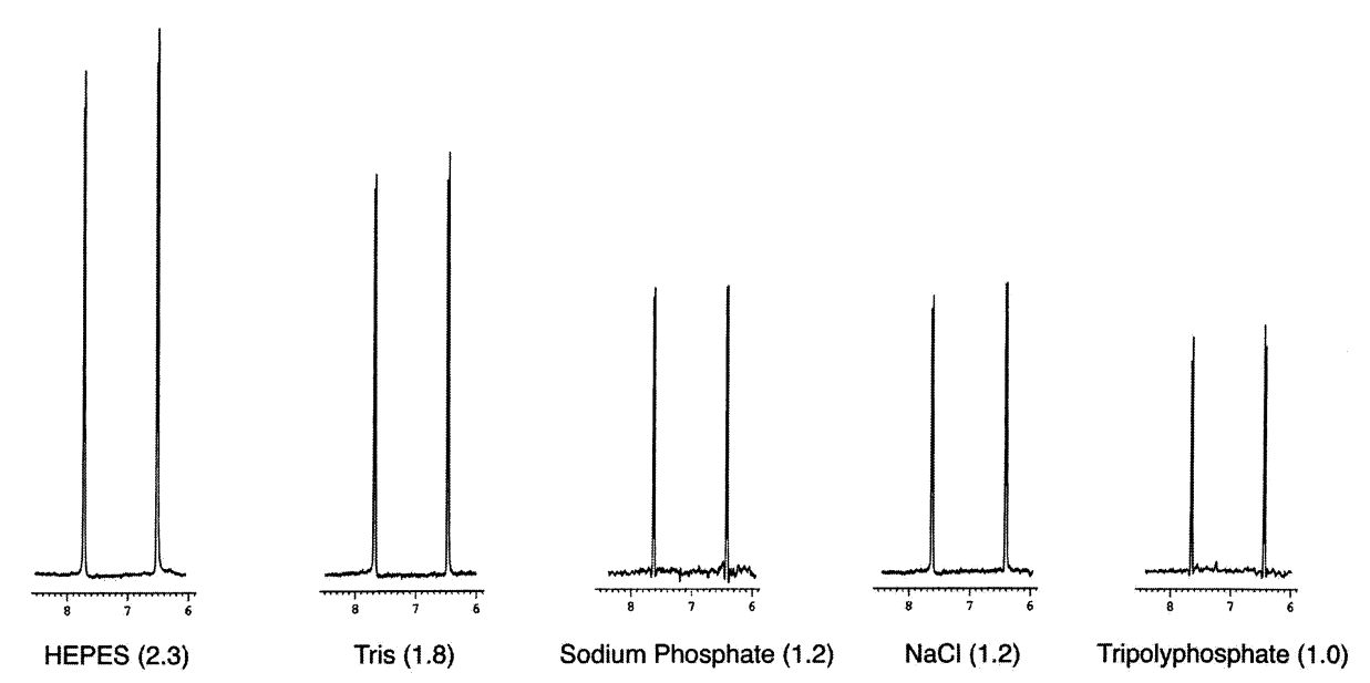
Sensitivities relative to tripolyphosphate of a 2 mM p-aminobenzoic acid sample dissolved in 200 mM buffer. Source: Kelly et al (2002)
Illustrated below is a similar effect on a biological sample (lysozyme in this case).
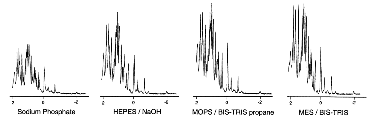
Source: Kelly et al (2002)
Therefore, time used to determine a good, low conductivity buffer suitable for the biological sample analyzed is always time well invested
Sample preparation
The sample has to be optically clear and free of particles in suspension. A 5 minute centrifugation, or a filtration through a 0.22 µm filter will remove any suspended matter.
On the day of the NMR experiment:
- Add D2O to a final concentration of 10 %
- Measure the pH of the sample and adjust if needed
- Centrifuge particles down from your sample and filter if needed
- Determine the exact sample concentration
- Transfer the sample to a clean NMR tube
- Label your NMR tube appropriately
Summary
- Aim for 500 µM of sample concentration in 500-550 µl (\~250 nmol of sample) if using a 5 mm NMR tube
- Avoid high salt concentrations, or look into alternative, low conductivity, buffers
- If you do not have enough material, consider using a 3 mm tube
Once you know your sample meets the above NMR requirements, you can reserve some time on the instrument by contacting me by email (normand.cyr@umontreal.ca).