Versions Compared

Key

  • This line was added.
  • This line was removed.
  • Formatting was changed.

What is photo-toxicity?

Under appropriate conditions of temperature (37°C), high humidity

...

, and a controlled atmosphere (5%

...

CO2), it is possible to grow cells in vitro.

...

Image Removed

Healthy cells

...

However, observing these cells under

...

a microscope can have unintended effects. This situation is

...

analogous to

...

exposing yourself to

...

sunlight: if the sunlight is too intense or the exposure lasts too long, you may suffer a sunburn. Similarly, cells can be affected by prolonged or intense light exposure during microscopy. While transmitted light (bright-field, phase-contrast, DIC) generally does not

...

interfere with cell biology, fluorescence excitation light can

...

cause significant phototoxic effects.

Image Added

Healthy cells

What does phototoxicity

...

look like?

...

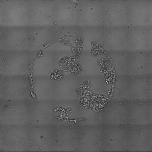
Prolonged exposure to intense illumination can cause significant cellular damage, resulting in retraction, detachment, and eventual cell death. The example below demonstrates this process (click on the image to watch the movie).

Image Added

Click on the image to see the movie

...


Cells subjected to 1 second of

...

70 mW, 550 nm light every 5 minutes. The total duration of the movie is

...

approximately 3 hours, during which the cells gradually retract, detach, and eventually die

...

Image Removed

Image AddedClick on the image to see the movie

...

Cells subjected to 1 second of

...

59 mW, 395 nm light and 1 s 750 mW 550nm every 5 minutes. The total duration

...

Image Removed

...

of the movie is approximately 8 hours. 

How can I identify

...

if phototoxicity has occurred?

The

...

most effective way to

...

detect phototoxicity is to

...

capture a larger field-of-view image after your acquisition.

...

Since phototoxicity is confined to the illuminated area,

...

comparing adjacent non-illuminated cells with those in the exposed region offers a good—though not perfect—approach.

By stepping back, you can observe that the damage is limited to the illuminated area. However, it’s important to note that the recorded region may be smaller than the actual exposed area.

It’s also crucial to understand that this method is not a flawless

Effects of Photo-toxicityImage Removed

...

control. A more ideal control would

...

involve using a separate dish with cells maintained

...

under identical conditions but without illumination.

...

This is because, in the image above, we

...

cannot definitively conclude whether

...

the affected circular

...

region has no impact on

...

nearby cells.

...

It’s possible that cell death

...

in the illuminated

...

region may release molecules that

...

influence the surrounding

...

cells. Therefore, the

...

most reliable control would be a

...

completely separate dish with unexposed cells.

Effects of Photo-toxicityImage Added

6 x 6 Tiles around the image area. 

What are the important factors

...

to consider when discussing phototoxicity?

Several key factors influence phototoxicity:

  • Amount of light:

...

  • Strong illumination causes more damage compared to dimmer light.
  • Wavelength: The energy carried by

...

  • light depends on its wavelength. Shorter

...

  • wavelengths carry higher energy and

...

  • tend to be more harmful.
  • Illuminated area: Concentrating

...

  • the same amount of light

...

  • on a

...

  • smaller area

...

  • results in

...

  • more localized damage.

...

  • Duration of illumination:

...

  • Prolonged exposure (e.g., 1 second vs. 10 ms) increases the risk of damage.
  • Repetition of illumination: Frequent exposure (e.g., 10 ms pulses applied 20 times per minute) is

...

  • more

...

  • damaging than less frequent exposure (e.g., once per minute).

How can you detect phototoxicity?

Empirically, phototoxicity can be identified by observing cell behavior. If

...

cells are not dividing

...

, retracting, or detaching, it may indicate phototoxicity.

To assess phototoxicity:

  • Acquire

...

  • a larger field-of-view image of the recorded area to

...

  • ensure no phototoxicity has occurred and to evaluate photobleaching.

...

  • Use a power meter

...

  • to measure the energy your cells are

...

  • exposed to.

    ...

      1. Measure the power at the objective using your

    ...

      1. usual imaging settings.

    ...

      1. Record the value in mW (

    ...

      1. milliwatts = Joules/second).

    ...

      1. Divide this value by the field of view

    ...

      1. area (in cm²) to calculate the irradiance

    ...

      1. in mW/

    ...

      1. cm².

    ...

    How to determine the maximum acceptable irradiance:
    Finding an irradiance level that is stress-free

    ...

    for your cells is

    ...

    critical:

    1. Expose your cells continuously to a defined irradiance.
    2. Observe them over several hours. If they show no signs of phototoxicity, gradually increase the irradiance and repeat the observation.
    3. Identify the maximum continuous irradiance that does not cause damage.

    Keep in mind that this value provides a baseline. Since most experiments do not involve continuous exposure, it is possible to exceed this threshold briefly. However, doing so may induce temporary stress in the cells. It is up to you to decide whether this level of stress is acceptable for your specific experiment and whether it might interfere with the biological processes you are studying.

    Image AddedClick on the image to see the movie

    This will provide a good idea of how strong are your cells.

    Obviously there is some flexibility since you will likely not continuously expose your cells. So you may pass above the maximum acceptable continuous irradiance value, which will stress your cells but eventually they will recover. Only you can determine if this amount of stress is experimentally acceptable and not modify the biology of what you want to measure.

    ...

    Cells were imaged for 100ms with 70mW of 550nm light every 5 minutes. Cells are dividing faster than the effect of photo-toxicity that is occurring. Again here the best way to control is to take an overview image at the end of the acquisition and compare exposed cells to non-exposed cells.

    ...

    Image Removed

    ...